Difference between revisions of "Alzheimer dementia"
(→References) |
|||
| (9 intermediate revisions by the same user not shown) | |||
| Line 1: | Line 1: | ||
| − | + | ===Diagnostics=== | |
Clinical criteria for Alzheimer’s dementia (McKhann et al, 2011) | Clinical criteria for Alzheimer’s dementia (McKhann et al, 2011) | ||
| Line 75: | Line 75: | ||
| − | + | ===Variants=== | |
| + | There are four common clinical presentations in Alzheimer's disease (Salardini 2019) | ||
| − | - | + | - Amnestic: typical problems with episodic memory (other domains may be affected, most commonly visuospatial cognition such as the inability to navigate), early changes in mesial temporal lobe, |
| − | - [[Visual variant (posterior cortical atrophy)]] | + | - [[Visual variant (posterior cortical atrophy) Alzheimer dementia]]: visual perception is compromised in the absence of any ophthalmological problem, typically presents in mid-50's or early 60's, relative sparing of other domains not involved in vision, neuroimaging with occipito-temporal atrophy |
| − | - [[Logopenic variant primary progressive aphasia]] | + | - [[Logopenic variant primary progressive aphasia]]: impairment in phonemic lexicon, leading to impaired naming, hesitation of speech, and spelling changes; memory deficits and anxiety often coexist |
| − | - [[Executive variant or frontal AD]] | + | - [[Executive variant or frontal AD]]: executive dysfunction and behavioral symptoms, shares overlap with bvFTD |
'''Autosomal dominant Alzheimer disease''': ''PSEN1, PSEN 2,'' and ''APP'' genes are known autosomal dominant genes in AD - see [[Genetics of Alzheimer disease]] subsection | '''Autosomal dominant Alzheimer disease''': ''PSEN1, PSEN 2,'' and ''APP'' genes are known autosomal dominant genes in AD - see [[Genetics of Alzheimer disease]] subsection | ||
| − | [[Early | + | [[Early onset Alzheimer dementia]] |
| − | |||
| + | ===Imaging=== | ||
| + | |||
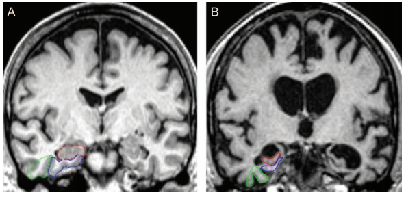
| + | [[File:Alzheimer's_dementia_coronal_MRI.png|none|frame|Comparison of normal (A) and severe atrophy (B) of medial temporal lobe. The hippocampus is traced in red, the entorhinal cortex in blue, and the perirhinal cortex in green. Severity of atrophy of the medial temporal lobe, including the entorhinal cortex and hippocampus, is predictive of future cognitive decline and conversion from MCI to AD (Duara et al, 2008)]] | ||
| + | |||
| + | |||
| + | ===Management=== | ||
| + | |||
| + | - acetylcholinesterase inhibitors (donepezil, galantamine, rivastigmine) | ||
| + | |||
| + | - memantine (moderate-severe stage) | ||
| + | |||
| + | - family support | ||
| + | |||
| + | - treatment of neuropsychiatric symptoms | ||
== References == | == References == | ||
| − | McKhann, G. M. et al. The diagnosis of dementia due to Alzheimer’s disease: Recommendations from the National Institute on Aging-Alzheimer’s Association workgroups on diagnostic guidelines for Alzheimer’s disease. Alzheimers Dement. 7, 263–269 (2011). https://pubmed.ncbi.nlm.nih.gov/21514250/ | + | Duara, R. et al. Medial temporal lobe atrophy on MRI scans and the diagnosis of Alzheimer disease. Neurology 71, 1986–1992 (2008). [https://pubmed.ncbi.nlm.nih.gov/19064880/ PubMed link] |
| + | |||
| + | McKhann, G. M. et al. The diagnosis of dementia due to Alzheimer’s disease: Recommendations from the National Institute on Aging-Alzheimer’s Association workgroups on diagnostic guidelines for Alzheimer’s disease. Alzheimers Dement. 7, 263–269 (2011). [https://pubmed.ncbi.nlm.nih.gov/21514250/ PubMed link] | ||
| − | Salardini, A. An Overview of Primary Dementias as Clinicopathological Entities. Semin. Neurol. 39, 153–166 (2019). https://pubmed.ncbi.nlm.nih.gov/30925609/ | + | Salardini, A. An Overview of Primary Dementias as Clinicopathological Entities. Semin. Neurol. 39, 153–166 (2019). [https://pubmed.ncbi.nlm.nih.gov/30925609/ PubMed link] |
Latest revision as of 20:27, 24 June 2021
Diagnostics
Clinical criteria for Alzheimer’s dementia (McKhann et al, 2011)
1) Probable Alzheimer’s disease dementia (core clinical criteria)
- a. Insidious onset over months to years
- b. Clear-cut history of worsening cognition by report or observation
- c. Initial and most prominent cognitive deficits on history and examination are one of the following:
- i. Amnestic presentation: impairment in learning and recall
- ii. Non-amnestic presentation
- 1. Language presentation: word-finding difficulties, deficits in other domains should be present
- 2. Visuospatial presentation: spatial cognition-object agnosia, facial recognition, simultagnosia, and alexia, deficits in other domains should be present
- 3. Executive dysfunction: impaired reasoning, judgment and problem solving, deficits in other domains should be present
- d. There is no evidence of
- i. Stroke temporally related to the onset of cognitive symptoms or presence of extensive infarcts or severe white matter hyperintensity burden
- ii. Core features of DLB other than dementia itself
- iii. Prominent features of bvFTD
- iv. Prominent features of semantic or non-fluent / agrammatic PPA
- v. Other active neurological disease, medical comorbidity, or use of medications with effects on cognition
2) Probable AD dementia with documented decline: core clinical criteria + evidence of decline on subsequent evaluation based on informants, formal neuropsychological evaluation, or standardized mental status examinations
3) Probable AD dementia in a carrier of a causative AD genetic mutation: core clinical criteria + presence of APP, PSEN1, or PSEN2 mutations
4) Probable AD dementia with evidence of AD pathophysiological process meets core clinical criteria + biomarker data
- a. High probability: amyloid PET or CSF + positive CSF tau, FDG-PET, or structural MRI
- b. Intermediate probability:
- i. unavailable, conflicting, or indeterminate amyloid PET or CSF + positive CSF tau, FDG-PET, or structural MRI
- ii. positive amyloid PET or CSF + unavailable, conflicting, or indeterminate CSF tau, FDG-PET, or structural MRI
- c. Uninformative: unavailable, conflicting, or indeterminate amyloid PET or CSF + unavailable, conflicting, or indeterminate CSF tau, FDG-PET, or structural MRI
5) Possible AD:
- a. Atypical: meets core clinical criteria for AD but either has a sudden onset or demonstrates insufficient historical detail or objective cognitive documentation or progressive decline
- b. Etiologically mixed presentation meets criteria for AD but has evidence of
- i. Stroke
- ii. Features of DLB other than dementia
- iii. Evidence of another neurological disease or medical condition with effects on cognition
6) Possible AD dementia with evidence of AD pathophysiological process: atypical clinical presentation plus the following biomarker data:
- a. High probability: positive amyloid PET or CSF + positive CSF tau, FDG-PET, or structural MRI
- b. Intermediate probability
- c. Uninformative: unavailable, conflicting, or indeterminate amyloid PET or CSF + unavailable, conflicting, or indeterminate CSF tau, FDG-PET, or structural MRI
Variants
There are four common clinical presentations in Alzheimer's disease (Salardini 2019)
- Amnestic: typical problems with episodic memory (other domains may be affected, most commonly visuospatial cognition such as the inability to navigate), early changes in mesial temporal lobe,
- Visual variant (posterior cortical atrophy) Alzheimer dementia: visual perception is compromised in the absence of any ophthalmological problem, typically presents in mid-50's or early 60's, relative sparing of other domains not involved in vision, neuroimaging with occipito-temporal atrophy
- Logopenic variant primary progressive aphasia: impairment in phonemic lexicon, leading to impaired naming, hesitation of speech, and spelling changes; memory deficits and anxiety often coexist
- Executive variant or frontal AD: executive dysfunction and behavioral symptoms, shares overlap with bvFTD
Autosomal dominant Alzheimer disease: PSEN1, PSEN 2, and APP genes are known autosomal dominant genes in AD - see Genetics of Alzheimer disease subsection
Early onset Alzheimer dementia
Imaging

Management
- acetylcholinesterase inhibitors (donepezil, galantamine, rivastigmine)
- memantine (moderate-severe stage)
- family support
- treatment of neuropsychiatric symptoms
References
Duara, R. et al. Medial temporal lobe atrophy on MRI scans and the diagnosis of Alzheimer disease. Neurology 71, 1986–1992 (2008). PubMed link
McKhann, G. M. et al. The diagnosis of dementia due to Alzheimer’s disease: Recommendations from the National Institute on Aging-Alzheimer’s Association workgroups on diagnostic guidelines for Alzheimer’s disease. Alzheimers Dement. 7, 263–269 (2011). PubMed link
Salardini, A. An Overview of Primary Dementias as Clinicopathological Entities. Semin. Neurol. 39, 153–166 (2019). PubMed link