File:Alzheimer's dementia coronal MRI.png
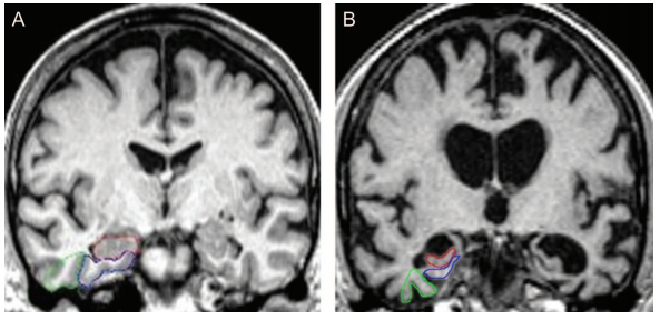
Revision as of 13:27, 14 June 2021 by Geoffrey (talk | contribs) (Comparison of normal (A) and severe atrophy (B) of medial temporal lobe. The hippocampus is traced in red, the entorhinal cortex in blue, and the perirhinal cortex in green. Severity of atrophy of the medial temporal lobe, including the entorhinal cort...)
No higher resolution available.
Alzheimer's_dementia_coronal_MRI.png (593 × 288 pixels, file size: 341 KB, MIME type: image/png)
Summary
Comparison of normal (A) and severe atrophy (B) of medial temporal lobe. The hippocampus is traced in red, the entorhinal cortex in blue, and the perirhinal cortex in green. Severity of atrophy of the medial temporal lobe, including the entorhinal cortex and hippocampus, is predictive of future cognitive decline and conversion from MCI to AD (Duara et al, 2008).
File history
Click on a date/time to view the file as it appeared at that time.
| Date/Time | Thumbnail | Dimensions | User | Comment | |
|---|---|---|---|---|---|
| current | 13:27, 14 June 2021 | | 593 × 288 (341 KB) | Geoffrey (talk | contribs) | Comparison of normal (A) and severe atrophy (B) of medial temporal lobe. The hippocampus is traced in red, the entorhinal cortex in blue, and the perirhinal cortex in green. Severity of atrophy of the medial temporal lobe, including the entorhinal cort... |
- You cannot overwrite this file.
File usage
The following page links to this file: